Since the original broadcast of this program, Taubenberger’s team has successfully created a genetic sequencing of the 1918 virus, resurrected the virus itself to study its effects on lung tissue, and this fall announced a striking similarity between the 1918 virus and today’s H5N1 avian flu virus. Their findings indicate that the 1918 virus originated as a bird flu, confirming the legitimacy of concerns about avian flu. The updated episode includes new material and interviews with Taubenberger that reflect these new findings.
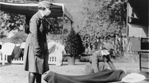
Over the centuries, humans have endured many influenza pandemics. Descriptions of the disease in Europe date back to 1100 A.D., and sporadic, isolated outbreaks have likely been occurring for many thousands of years. But no flu ever struck as fast, as hard, and with such lethal power as the 1918, or “Spanish Flu,” which rivals the Black Death as the deadliest epidemic in history. To a world already ravaged by war, the 1918 pandemic was crippling; some 30 to 40 million people died worldwide, with the highest death rate occuring in young men and women. In the United States, 675,000 people died, including over 40,000 G.I.s — 40 percent of all of those who perished in World War I.
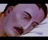
This illustration shows a British soldier displaying the distinctive blue coloration of heliotrope cyanosis.
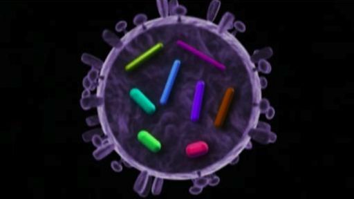
In 1918, scientists didn’t know that the culprit behind the carnage was a virus, a simple capsule filled with a few snips of genetic material that allow it to harness its host’s cellular machinery and make limitless copies of itself. In many ways, the 1918 bug — a variety of influenza A, the most common cause of flu in humans — was no different than any other influenza virus. Within the viral capsule are eight strands of RNA, which carry a total of just eight genes. Two of the genes produce the sugar-rich proteins hemagglutinin and neuraminidase, which stud the surface of the virus with knob-like protrusions. The virus gains entry into a host’s cells with the aid of these two proteins, which have a distinctive structure in every flu strain and are the target of both flu vaccines and the body’s immune response. To start the infection process, enzymes naturally present in the host cut the hemagglutinin protein into two pieces so that it can bind to a receptor on the cell’s surface. The virus is then enveloped, pulled into the cell, and eventually released and broken apart when the neuraminidase protein destroys the receptor that connects virus to cell.
The RNA strands move toward the cell nucleus. There, the cell reproduces each strand and creates new proteins that are then packaged into capsules and ultimately released from the cell to infect other cells. In humans, the influenza virus selectively attacks and destroys the cells that line the upper respiratory tract, bronchial tubes, and trachea. One to two days after infection, the symptoms appear: shaking chills, fatigue, muscle ache, listlessness, and high fever. Flu mortality rates typically linger around 0.1 percent (despite the widespread availability of flu vaccines and modern antiviral drugs, 36,000 people still die every year in the United States from complications of the flu); in the 1918 flu, the rate was twenty-five times higher, with deaths usually the result of secondary bacterial pneumonia or bronchitis. Many flu victims in 1918 displayed a distinctive dusky blue-gray pallor on their face, lips, and ears, called heliotrope cyanosis — a mark of a patient who is being suffocated to death by a buildup of fluid and cells in his lungs.